Measurement and testing
Published over 4 years ago. See the latest and most current information on Measurement and testing.
From high-performance synthetic plastics to natural substances like wool, silk and cellulose, microscopic polymer characterisation is used to analyse a wide range of materials. The technique reveals valuable data about the surface structures of polymers and the individual atoms that make up the unique materials.
Analysts rely on a variety of techniques to observe polymer materials and generate high-resolution images. We take a closer look at some of the most useful methods below:
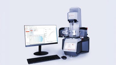
Scanning Electron Microscopy uses a focused beam of electrons to scan the surface of a polymer material. As electrons hit the surface they interact with atoms and generate distinctive signals. Data is used to analyse the topography of the sample and observe any unique textures and surface patterns.
SEM is also used to analyse morphological properties, aka the size, shape and arrangement of particles. Information about elemental composition and crystalline structure can also be revealed using Scanning Electron Microscopy. The technique is fast and simple, with minimal sample preparation and good compatibility with most polymer materials.
Transmission Electron Microscopy relies on specialised instruments to analyse polymers. The microscopes generate high-resolution images of samples using a particle beam of electrons. With the ability to magnify objects by up to two million times, TEM is one of the most advanced microscopic polymer characterisation techniques available to scientists.
Recently, High-Resolution Transmission Electron Microscopy (HRTEM) has pushed the limits of TEM. The cutting-edge technique has transformed the polymer science field and allowed analysts to generate incredibly detailed images of molecular structures. A recent article published in the journal Nature Communications explored the use of antioxidants as a way to reduce the risk of beam damage caused by HRTEM analysis.
“Characterising the effects of beam damage by calculating critical dose DC values from the decay of electron diffraction peaks shows that beam damage of conjugated polymers in the TEM can be minimised by using antioxidants at room temperature, even if the antioxidant does not alter or incorporate into polymer crystals,” reads the abstract.
SPM uses a physical probe to scan and map the surfaces of polymer materials. Interactions between the probe and the surface are recorded and used to reveal the topographic properties of the sample. Analysing the performance and characteristics of the thin, transparent polymer films used for food packaging is one application for Scanning Probe Microscopy.
Microscopic polymer characterisation is suitable for many applications but it’s not the only analytical tool available to scientists. Find out more about other techniques, including chromatographic polymer characterisation used to map the composition and molecular weight distributions of polymers, in ‘Polymer Characterisation - Techniques, Types & Properties.’
PIN 27.3 June/July 2026