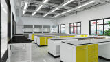
Laboratory products
Research into the world’s most dangerous pathogens demands high-containment laboratory facilities. Whether the organism under study is an emerging virus or a multidrug-resistant bacterium, any pathogen classified at Containment Level 3 or above must be handled inside biosafety cabinets within purpose-built laboratories. These essential safety measures protect researchers and the wider community, but they also impose significant practical constraints. The physical space inside a biosafety cabinet is limited, decontamination protocols are rigorous, and every piece of equipment introduced into the contained environment must be compatible with these restrictions. As a result, many standard laboratory techniques that are routine at lower containment levels become difficult or impossible to perform when working with live hazardous organisms.
This has long been a bottleneck in infectious disease research. Real-time, quantitative assays for tracking pathogen growth that make the foundation of studies into virulence, drug susceptibility, and host–pathogen interactions often rely on instruments that are simply too large to fit inside a biosafety cabinet. The consequence is that researchers must either work with killed or inactivated organisms, sacrificing biological relevance, or remove samples from containment for analysis, introducing logistical complexity and safety concerns. A new generation of compact luminometers is now changing this equation, enabling live-pathogen assays to be conducted entirely within the biosafety cabinet.
One research group at the forefront of this approach is based at the University of Surrey, where Professor Rachel Simmonds leads a team investigating Buruli ulcer, a neglected tropical disease caused by Mycobacterium ulcerans. Buruli ulcer predominantly affects rural communities in West and Central Africa, South-East Asia, and parts of Australia, with around 2,000 cases reported annually. The disease causes progressive damage to skin and soft tissue, often leading to disfigurement or amputation. Despite this severe clinical impact, Buruli ulcer remains one of the least understood mycobacterial diseases, partly because M. ulcerans is exceptionally difficult to work with under the containment conditions it requires.
Professor Simmonds’ group is tackling a fundamental question in Buruli ulcer pathogenesis: can M. ulcerans replicate inside macrophages? Macrophages are phagocytic immune cells that normally engulf and destroy invading pathogens, yet several mycobacterial species, most notably M. tuberculosis, have evolved strategies to survive and persist within them. Whether M. ulcerans similarly exploits macrophages as a replicative niche has remained an open question.
“We are starting to realise the importance of macrophages in the pathogenesis of the disease,” explained Kwabena Boateng, a PhD student in Simmonds’ group. “One of the questions that we want to answer is whether the bacteria can actually grow inside macrophages.”
Answering this question requires the ability to track bacterial growth within host cells over time. Traditional methods for quantifying microbial growth, such as colony-forming unit counts, are time-consuming and labour-intensive. Microscopy-based counting becomes impractical when all manipulations must be performed within the confined space of a biosafety cabinet. Flow cytometry and conventional plate readers, while powerful, require bench-top instruments that are too large to operate inside the cabinet. These constraints apply equally whether the pathogen in question is a bacterium, a virus, or a fungus; containment imposes the same physical limitations regardless of the organism.
Bioluminescence-based assays offer a compelling solution. The principle is well established: an organism is engineered to express a luciferase enzyme, which catalyses a light-emitting reaction. As the pathogen replicates, luciferase production increases proportionally, and the resulting light output serves as a read-out for growth. The technique has previously been applied to studies of bacterial physiology, antibiotic susceptibility screening, and viral replication kinetics. In virology, for example, recombinant reporter viruses expressing luciferase are widely used to quantify viral entry, replication, and the efficacy of antiviral compounds in cell-based assays. The same principle applies to intracellular bacteria and parasites, where luminescence can report on pathogen behaviour within host cells without the need to lyse or fix them.
The advantages over alternative quantification methods are considerable. Luminescence does not cause phototoxicity or suffer from photobleaching, meaning the same culture can be measured repeatedly to generate continuous growth curves. It is highly sensitive, capable of detecting small changes in pathogen number that might be missed by other methods. Crucially, it requires minimal sample handling, reducing both contamination risk and the exposure of laboratory workers to hazardous material.
The development of compact luminometers, such as MyGlo Reagent Reader (Promega Corp), has enabled luminescent-based assays to be used in high-containment environments. These instruments have a footprint similar to a standard 96-well microplate, with minimal cabling, allowing them to sit comfortably inside the working area of a Class II or Class III biological safety cabinet. Data are transmitted to a computer outside the cabinet, so the entire measurement process takes place within containment.
This design philosophy addresses the core challenge of high-containment research: enabling sophisticated analytical techniques without compromising biosafety. The small footprint also means the instrument can remain inside the cabinet between readings, avoiding the need for repeated decontamination cycles that would be required if equipment were moved in and out. For longitudinal studies requiring measurements over days or weeks, as is common with slow-growing pathogens like mycobacteria, this practical advantage is substantial.
For the Surrey team, this capability transformed what was experimentally possible. Professor Simmonds recognised the potential immediately. “It’s very small,” she noted. “It’s not much bigger than the 96-well plate and has a single wire connected to it, and I could see immediately that it would work very well inside the cabinet in a high-containment environment.”
The group engineered M. ulcerans to express a luminescent reporter by cloning a luciferase-encoding operon into the bacterium. They then developed a protocol in which macrophages are infected with the luminescent strain in 96-well plates, and the plates are read at regular intervals using the compact luminometer inside the biosafety cabinet. Changes in luminescence over time reveal whether the bacteria are replicating, persisting, or being killed within the host cells.
“We infect the macrophages with these luminescent bacteria and we can track the increase in luminescence over time, which we use as a correlative marker for growth,” explained Boateng.
While the Buruli ulcer work provides a compelling example, the same approach has broad applicability across infectious disease research. Any pathogen that can be engineered to express a luminescent reporter, or for which luminescent reporter assays already exist, can potentially be studied using luminometry within containment. In virology, this is particularly relevant for work with high-consequence viruses at Containment Level 3 and above, where reporter virus systems are already established tools. Luminescent reporter assays for influenza, coronaviruses, and other respiratory pathogens could be conducted entirely within the biosafety cabinet, enabling real-time antiviral screening and viral growth kinetics studies under conditions that more accurately reflect genuine containment workflows.
The work at the University of Surrey illustrates how advances in instrumentation, and reducing the size of a luminometer to fit inside a biosafety cabinet, can have outsized implications for research into some of the world’s most challenging infectious diseases. Utilising luminescent workflows within biosafety cabinets represents a shift in what is experimentally achievable under high-containment conditions.
As the infectious disease research community continues to grapple with emerging pathogens, antimicrobial resistance, and neglected tropical diseases, tools that bring sophisticated real-time analytics directly into the containment environment will play an increasingly important role. The principle is simple but powerful: the most informative experiments on dangerous pathogens are those conducted on living organisms, and the instrumentation must go where the science needs to happen.
Joanna JS Butler, Roderick Hay, Richard O Phillips, Caroline Erolin and Rachel E Simmonds.
‘Medical illustrations of Buruli Ulcer’ Available from the University of Surrey’s open research repository
PIN 27.2 Apr/May 2026


.jpg)